

Chloe is involved in TWO OVC Clinical Trials!
Study Investigator: Dr. Michelle Oblak
Study Title: Evaluation of Sentinel Lymph Node Mapping in Dogs with Lung Tumours Using CT Lymphography and Intraoperative Indocyanine Green
AND
Study Investigators: Dr. Brenda Coomber and Dr. Paul Woods
Study Title: Collection of Biological Specimens from Dogs Scheduled for Biopsy or Surgery for Suspected or Known Cancer
Generously Supported by: PetDx
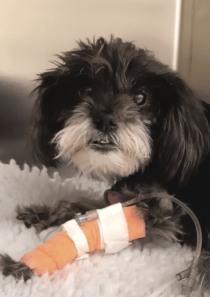
Chloe was diagnosed with pulmonary adenocarcinoma (a tumour originating from her lung). Part of cancer staging includes diagnostic imaging such as CT, MRI, ultrasound or x-rays. A lung lobectomy (surgical removal of the tumour and part of the lung) was recommended for Chloe’s treatment.
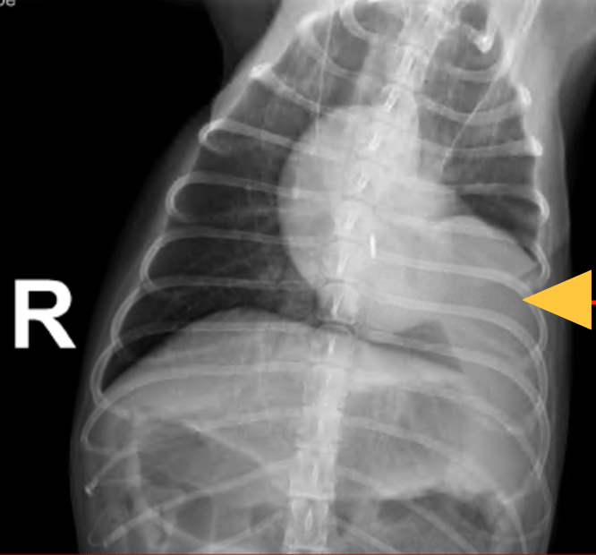
This is an x-ray of Chloe’s chest. Her tumour is indicated by the arrow.
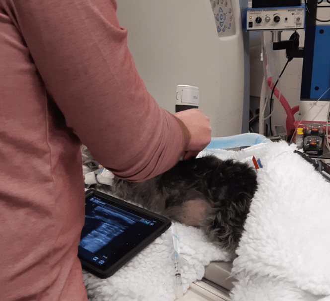
During her CT, an ultrasound was used to sample the tumour for diagnosis (fine needle aspirate) and to inject the a contrast dye for lymph node evaluation. This allows for preoperative tumour staging, essential to understanding tumour metastasis.
During surgery, Dr. Oblak used a special dye called indocyanine green (ICG) and a near-infrared fluorescence (NIRF) camera is used to make the tumour and lymph nodes ‘glow-in-the-dark.’ Typically, the neighbouring lymph node(s) may not have been removed as they can be hard to find but because of the ‘glow,’ these were easily identified. The lung mass and lymph node were sent to the pathologist who confirmed that that the lymph node was in fact positive for metastatic disease.
By using this new technology, Dr. Oblak can make sure that her patients have the best possible staging and treatment outcomes! Chloe is doing very well and is continuing treatment with chemotherapy from the OVC Oncology service.
By participating in this interventional study, Chloe is helping us to develop protocols to better diagnose and treat dogs with lung tumours in the future. The results can also be applied to other tumour types in both dogs and cats! Dr. Oblak is also working closely with human researchers that hope to apply these techniques for surgery in humans with lung tumours!