

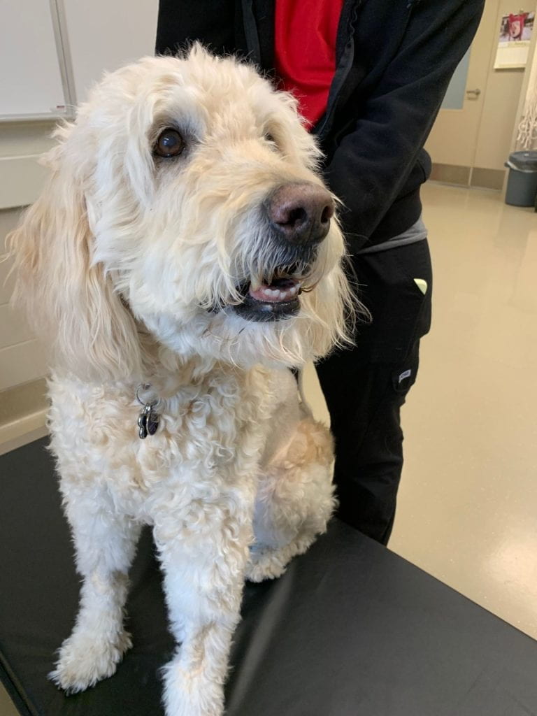
Monty is involved in two OVC Clinical Trials!
Study Investigator: Dr. Michelle Oblak
Study Title: Evaluation of Sentinel Lymph Node Mapping in Dogs with Lung Tumours Using CT Lymphography and Intraoperative Indocyanine Green
Generously Supported by: OVC Pet Trust and the AHP Research Chair
AND
Study Investigators: Dr. Brenda Coomber and Dr. Paul Woods
Study Title: Collection of Biological Specimens from Dogs Scheduled for Biopsy or Surgery for Suspected or Known Cancer
Generously Supported by: PetDx
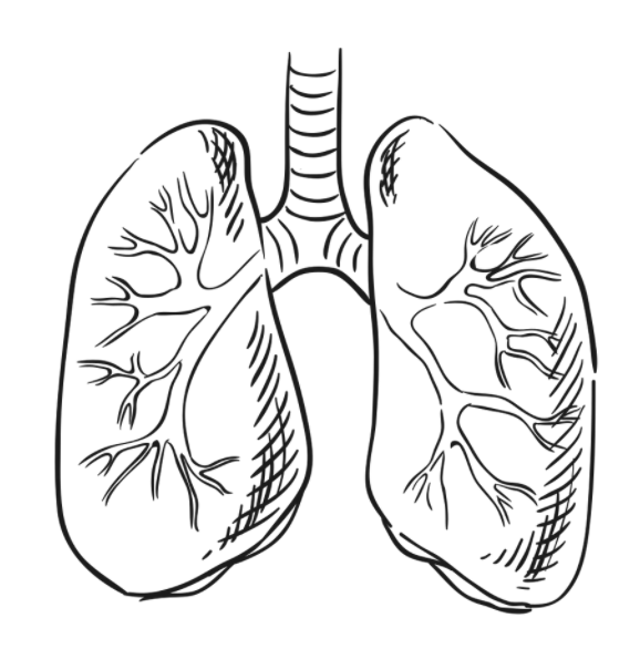
Monty was diagnosed with pulmonary adenocarcinoma (a tumour in his left lung). He did not have any visible metastases!
A lung lobectomy (surgical removal of the tumour and part of the lung) followed by chemotherapy was recommended for Monty’s treatment.
He was able to participate in 2 clinical studies. By participating in the ICG lung trial, Monty is helping to develop protocols to better diagnose and treat future dogs with lung tumours!
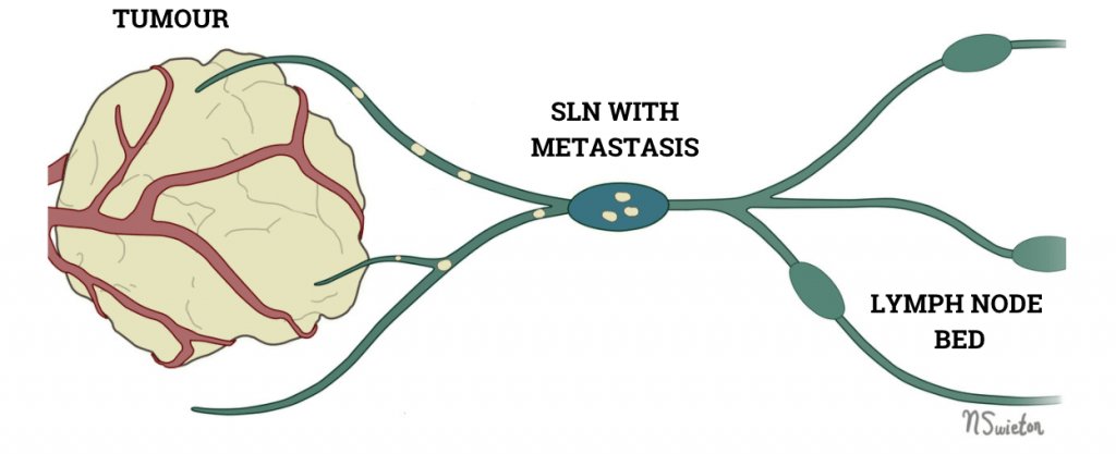
For many cancers, the first place of spread (metastasis) is to the local lymph nodes.
The first lymph node that the tumour drains is called the sentinel lymph node (SLN).
You might expect that the SLN would be the first lymph node downstream from the tumour, but this is not always true. By mapping the SLN, we identify and assess these draining lymph nodes. By removing the SLN, rather than the entire lymph node bed, we can decrease the complications after surgery and provide the most accurate information about the cancer’s metastasis.
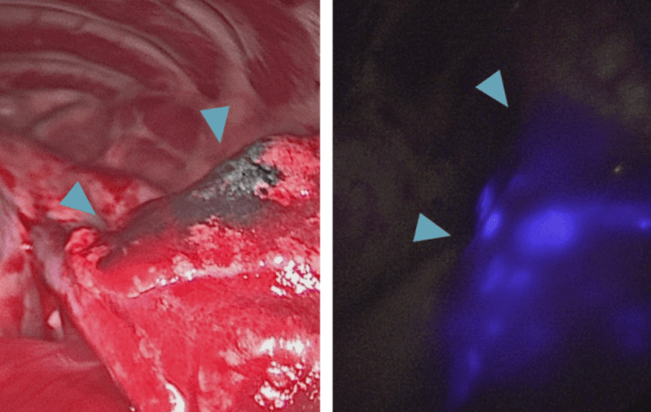
During surgery, a special dye called indocyanine green (ICG) and a near-infrared fluorescence (NIRF) camera are used to make the tumour and lymph nodes ‘glow-in-the-dark‘. By comparing what we can see with visible light to what glows under NIRF, Dr. Oblak can make sure Monty has the best possible surgical outcome!
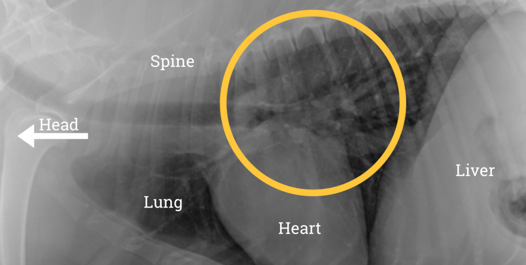
Monty’s lung tumour is indicated by the arrows.
Monty is doing well and is continuing his treatment with chemotherapy from the OVC Oncology service.