


Jax is involved in TWO OVC Clinical Trials!
Study Investigators: Dr. Owen Skinner (MUCVM) and Dr. Michelle Oblak (OVC)
Study Title: Evaluation of Regional Lymph Node Metastasis in Canine Thyroid Carcinoma
Generously Supported by: Clinician Scientist Grant
AND
Study Investigators: Dr. Brenda Coomber and Dr. Paul Woods
Study Title: Collection of Biological Specimens from Dogs Scheduled for Biopsy or Surgery for Suspected or Known Cancer
Generously Supported by: PetDx
Jax was diagnosed with thyroid carcinoma. Thyroid tumours are the most common cancer of tissues that produce hormones in dogs.
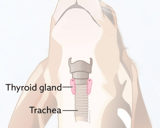
In dogs, the thyroid glands are located at the base of the neck on either side of their windpipe (or trachea).
This cancer can spread locally into the windpipe and esophagus but can also metastasize to other parts of the body including the lymph nodes and lungs. Surgical removal of the thyroid gland, when possible, is the recommended treatment for this disease. For this study, the neighbouring lymph nodes to the tumour were also removed.
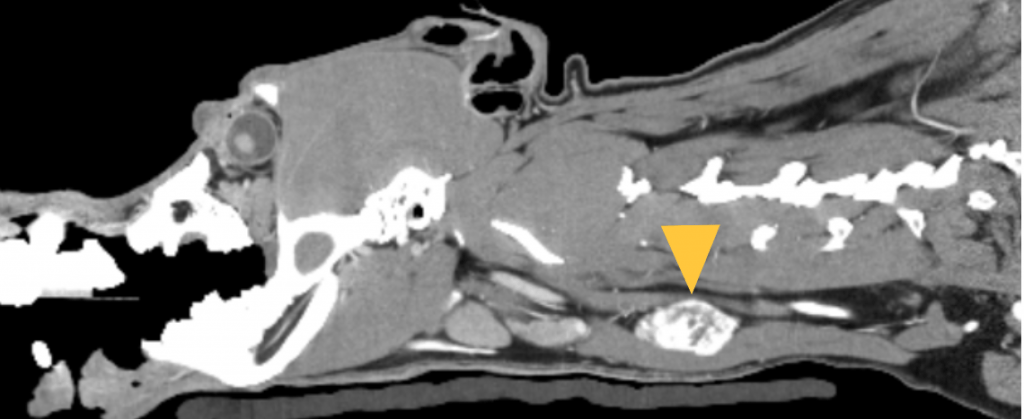
Prior to surgery, Jax had a CT scan of his head and neck to assess his tumour and lymph nodes for any signs of metastases. A CT scanner uses x-rays to produce images of the underlying tissues (muscles, organs and glands like the thyroid). The arrow indicates Jax’s tumour. This is a sagittal view; looking right through the side of his head and neck!
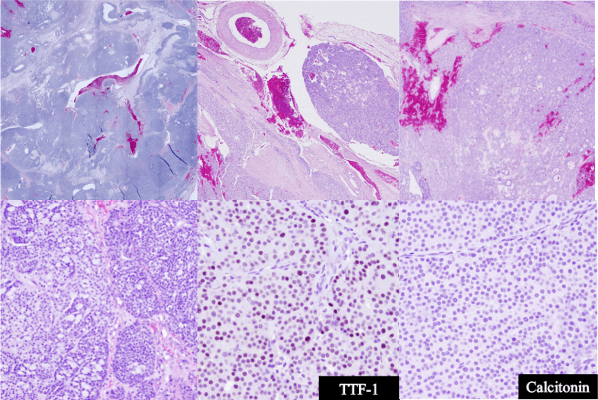
The removed tissues were shipped to Dr. Skinner’s team at the Missouri Veterinary Health Centre for a detailed assessment. Using special stains, we can identify cancer deposits that may not have been seen using traditional staining techniques! It can also help differentiate between the 4 subtypes of thyroid carcinoma: follicular, medullary, compact and mixed. Staining allows us to view the cellular structures, organization of cells and any abnormalities within the tissues.
These are highly magnified images of Jax’s thyroid gland. Based on the cellular staining, he has the ‘mixed’ cancer subtype (tumour cells in both follicles and packets).
Jax is doing well with no signs of recurrence of his cancer. He will return for follow-up appointments with the OVC Oncology service.
By participating in this collaborative study between the University of Missouri Veterinary Health Centre and University of Guelph Ontario Veterinary College, Jax is helping us learn more about thyroid cancer and how it spreads to the lymph nodes. Collected samples provide valuable data about the subtypes of thyroid carcinoma and if subtypes have an effect on prognosis. We can also assess the accuracy of CT imaging for confirmation of lymph node involvement.
The goals of both studies that Jax has enrolled in include developing earlier detection methods for cancer in our companion animals!