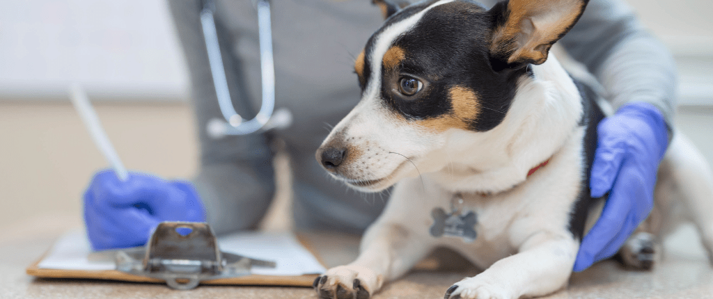
a white jack Russell terrier with black and brown spots laying on a desk as a woman wearing purple gloves and a stethoscope holds the dog in place while writing on a clipboard.

All Active Canine Clinical Trials

New studies are being added all the time – check back soon if you don’t see one available for your dog!

Cardiology


A brown dog is laying down next to a glass bowl of kibble.

Evaluating Grain-Free Diets and Cardiac Function in Dogs

Scientific Title: Plasma Metabolomic Profiles and Owner Feeding Practices in Dogs with Systolic Dysfunction or Dilated Cardiomyopathy

Study Investigator: Dr. Shari Raheb

Graduate Student: Sydney Banton (PhD)

Dilated cardiomyopathy (DCM) is a common heart disease in dogs, often caused by genetics. Recently, there has been concern about grain-free dog diets possibly contributing to DCM, but the majority of dogs eating these diets are still healthy. It is important to understand why some dogs may develop heart problems while eating grain-free diets and other dogs do not.

Inclusion criteria:

  • Two groups of dogs will be included in this study:
    • Healthy dogs >1 year of age that have been eating the same grain-free kibble diet for ≥ 6 months
    • Dogs with suspected cardiac dysfunction (and no additional comorbidities) that have been eating the same grain-free kibble diet ≥ 6 months
  • Dogs will be ineligible if they are receiving cardiac medication or their diet has been recently changed.
  • Breeds including Doberman, Boxer, Irish Wolfhound, Newfoundland, Great Dane, Cocker Spaniel and Portuguese Water Dog or mixes of these breeds will be ineligible to participate.


Diagnostic Imaging


ON HOLD FOR RECRUITMENT – Evaluating the Effect of Minimally Invasive Surgery on the Intracranial Pressure in Dogs

Scientific Title: The Effect of Laparoscopy on Intracranial Pressure as Measured by Optic Nerve Sheath Diameter

Study Investigator: Dr. Ryan Appleby

Graduate Student: Dr. Kevin Mitchell (DVSc)

Minimally invasive techniques like laparoscopy offer benefits such as less pain and quicker recovery, but they can also increase pressure inside the skull. This pressure change has been studied in humans but not in dogs. The optical nerve sheath contains cerebral spinal fluid and when intracranial pressure increases, the walls of the sheath expand. Ultrasound can be used to measure the changes in the optic nerve sheath diameter (ONSD). Understanding how intracranial pressure changes affect dogs could help with anesthesia and surgery planning for laparoscopic procedures in veterinary medicine.

Inclusion Criteria:

  • Healthy female dogs (>10kg) undergoing an open spay
  • Healthy dogs (>10kg) undergoing a laproscopic spay or spay and gastropexy

Emergency and Critical Care


Internal Medicine


Small while and black dog (Yorkie) looks up at the camera.

How Well Do Dissolvable Capsule Cameras Work in Small Dogs for Gut Imaging?

Study Investigator: Dr. Alice Defarges

Capsule endoscopy has revolutionized the approach of endoscopy in humans and dogs, allowing for the evaluation of the entire gut in a minimally invasive way, without the need for general anesthesia. Dissolvable non-endoscopic capsules are commonly used in medium to large breed dogs for gut imaging. The utility and speed at which the capsule passes through the gut of small breed dogs has not been evaluated.

Inclusion criteria:

  • Healthy dogs between 3 and 8 kg, receiving no medication affecting gut motility (i.e. prokinetics or opioids), no vomiting, diarrhea or regurgitations in the last month

Image of brown and black pug walking with tongue out.

ON HOLD FOR RECRUITMENT – Evaluating the Impact of Elective Surgery on the Gut Bacteria of Dogs

Scientific Title: Impact of Elective Orthopedic Surgery on the Fecal Microbiome of Dogs

Study Investigator: Dr. Shauna Blois

The gut microbiome is a collection of bacteria and other microorganisms living within the gastrointestinal tract, including many beneficial bacteria. When dogs have surgery, this may change the microbiome population and these changes could have an impact on a dog’s overall health.

Inclusion criteria:

  • Dogs undergoing elective orthopedic surgery (e.g., TPLO, etc) at OVC, with no newly diagnosed conditions within 1 month of surgery

Neurology


Image of a light brown french bulldog holding a grey teddy bear in its front paws

Understanding Spinal Muscle Function in Dogs with Intervertebral Disc Disorders

Scientific Title: Canine Paraspinal Muscle Biomechanical and Physiological Properties Related to Intervertebral Disc Disorders

Study Investigator: Drs Alex Chan, Fiona James and Stephen Brown

Graduate Student: Josh Briar (PhD)

Spinal degeneration causes pain and disability in both dogs and humans with treatment often involving surgery. Because spinal degeneration is associated with the degeneration and dysfunction of spinal muscles, these muscles are a prime target for patient rehabilitation strategies. In humans and dogs, we do not fully understand how these muscles degenerate and the impact of these changes on muscle function. The research team is working closely with human clinicians and recruiting both humans and dogs into this comparative study!

Inclusion criteria:

  • Dogs with a confirmed diagnosis of acute disk herniation and/or intervertebral disc degeneration (IVDD) undergoing MRI and standard of care surgery

Image of two brown dogs (one spotted brown) sitting outside

Are These Seizures in Dogs?

Study Investigator: Dr. Fiona James

Graduate Student: Charly McKenna (PhD)

Seizures are common in veterinary medicine, and epilepsy is the most common brain disease in dogs. Diagnosing epilepsy can be tricky because there are many different kinds of seizures and other disorders might look like epilepsy. It’s important to know if a dog’s episodes are actually seizures, as they might need treatment. Using video recording and electroencephalography (EEG), we can confirm if seizure activity is happening and classify the type of seizure.

Inclusion criteria:

  • Dogs that are between 6 months and 6 years of age that experience episodes similar to seizures

Image of 4 border collies (One back and white, two black white and brown, one brown and white) laying outside

Do Anti-Seizure Drugs Work in Dogs?

Study Investigator: Dr. Fiona James

Graduate Student: Charly McKenna (PhD)

Even with anti-seizure drug (ASD) treatment, a proportion of dogs may continue to have seizures or experience intolerable side effects from their medication. Accurate seizure control impacts the quality of life and survival in epileptic dogs and also their caretaker’s quality of life.

Inclusion criteria:

  • Dogs between 6 months and 6 years of age with normal neurologic exam and at least one year since index seizure (known as Tier I idiopathic epilepsy)
  • Any dog with Tier II idiopathic epilepsy regardless of age, neurologic exam status, or time since the first onset of seizures (must have normal MRI and CSF & bloodwork)
  • Dog’s veterinarian is planning to add a new ASD regardless of previous ASD status

Image of two husky dogs (one black and brown and one white and brown) sitting outside in the snow

ON HOLD FOR RECRUITMENT – Investigating Canine Behaviour using Wearable Biomonitors

Scientific Title: Pilot study of wearable biomonitors to explore the behavioural and environmental context of canine seizures

Study Investigator: Dr. Fiona James

Idiopathic epilepsy (IE) is the most common neurological disorder diagnosed in dogs. IE has a significant negative impact on affected dogs and their owners’ quality of life. Through the use of wearable technologies for both pets and owners, we can increase our understanding of epileptic canine behaviour and gain insight to better predict future seizure occurrence.

Inclusion criteria:

  • Dogs (Golden retrievers, Labrador retrievers, Huskies, German Shepherds, Border Collies, Australian Shepherds, Standard Poodles, and mixes of these breeds) between the ages of 2-7 years old
  • Two groups:
    • Dogs with Tier I idiopathic epilepsy diagnosis with no other health concerns/underlying conditions
    • Neurotypical with no other health concerns/underlying conditions

Image of a chocolate brown labrador outside with his tongue out

Evaluating the Structural Brain Differences of Dogs Diagnosed with Idiopathic Epilepsy

Scientific Title: Craniocerebral Topographical Mapping in Dogs with Idiopathic Epilepsy

Study Investigator: Dr. Fiona James

Graduate Student: Grace Kadler (PhD)

Diffusion tensor imaging (DTI) is sensitive to the molecular movement of water and can provide information on the integrity and pathology of the brain on a cellular level. In dogs with idiopathic
epilepsy (IE), the microstructures within the brain may be compromised. In order to better understand the abnormal brain structures associated with IE and
potentially improve diagnostic and treatment options, we first need to compare DTI between IE and neurotypical dogs.

Inclusion criteria:

  • All dogs must be mesocephalic (have an average muzzle length) and undergo a MRI at the Ontario Veterinary College
    • Neurotypical dogs with no obvious structural brain abnormalities and/or neurological disorders
    • Dogs with no physical head abnormalities with a planned EEG recording
    • Dogs diagnosed with generalized IE that have no other medical conditions

Image of a black and brown dog outside with orange leaves

Investigating the Placement of EEG Electrodes in Dogs with Epilepsy Using 3D Reconstruction

Scientific Title: Craniocerebral Topographical Mapping for Improved Canine Electroencephalographic (EEG) Lesion Localization

Study Investigator: Dr. Fiona James

Graduate Student: Dr. Stephen Everest (DVSc)

To improve the diagnosis and characterization of canine epilepsy, we need to better understand how our scalp electrodes for electroencephalography (EEG) map to the underlying brain surface and establish a best practice for electrode placement and subsequent seizure detection.

Inclusion criteria:

  • All dogs must be mesocephalic (have an average muzzle length) and undergo a MRI and CT at the Ontario Veterinary College
    • Neurotypical dogs with no obvious structural brain abnormalities and/or neurological disorders
    • Dogs with no physical head abnormalities with a planned EEG recording
    • Dogs diagnosed with generalized IE that have no other medical conditions

Oncology


Evaluating Palladia® for the Treatment of Canine Oral Melanoma

Study Investigator: Dr. Anthony Mutsaers

Graduate Student: Dr. Gabby Allegrini (DVSc)

Palladia is a type of drug originally used to treat recurrent mast cell tumours in dogs. New evidence has shown that Palladia may be useful for treating other cancer types. Oral melanoma is a locally invasive and highly metastatic cancer. In some cases, even with aggressive therapy, survival times can be as little as 6 months. The use of Palladia, if other treatments have failed, or if traditional therapy is declined, may extend survival in these patients.

Inclusion criteria:

  • ​Dogs with a confirmed diagnosis of oral melanoma through biopsy and measurable disease
  • Dogs that have previously received the melanoma vaccine will still be eligible

Image of a brown and white boxer dog with its paw in the air being held by a human hand.

Evaluating Blood Markers in Dogs with Mast Cell Tumours

Scientific Title: Micro RNA Profiling for Diagnosis and Prognosis in Canine Mast Cell Tumours

Study Investigator: Dr. Darren Wood

Graduate Student: James Mori (MSc)

Mast Cell tumours (MCT) are a type of cancer that arises from mast cells and is a relatively common diagnosis in dogs. There are many different subtypes of MCT which can impact how well dogs will respond to treatment and their overall prognosis. MicroRNA (miRNA) can be found in the blood and these small molecules regulate a number of cellular activities including cell growth, differentiation, development, and apoptosis. The use of biomarkers such as miRNA for cancer diagnosis and/or prognosis has high potential in veterinary medicine.

Inclusion criteria:

  • ​Dogs with naturally occurring mast cell tumours undergoing standard of care surgical excision
  • Dogs must not have a previous history of surgical removal with microscopic analysis, prior therapy (ie: chemotherapy or radiation) and/or active disease unrelated to the tumour

Image of a grey and white sheepdog laying outside with its tongue out.

Evaluating the Use of Fluorescent Dyes in Dogs Undergoing Liver Mass Removal Surgery

Scientific Title: Evaluating the Use of Fluorescent Dyes in Dogs Undergoing Liver Mass Removal Surgery

Study Investigator: Dr. Brigitte Brisson

Graduate Student: Dr. Rachel Dobberstein (DVSc)

Dogs are prone to developing liver cancer. In humans, fluorescent dyes are used to identify liver tumours and determine appropriate tissue margins for complete surgical removal. This same technique may be used in dogs with liver tumours and as it does in people, the fluorescence imaging may guide surgeons in determining the required margins to remove the entire tumour.

Inclusion criteria:

  • ​Dogs undergoing an open (laparotomy) abdominal surgery at OVC-HSC
  • Dogs diagnosed with primary liver neoplasia or abdominal neoplastic lesions that commonly metastasize to the liver by time of diagnosis e.g. splenic HSA.
  • Cancer diagnosis will be suspected based on recent (<14 days) abdominal ultrasound or abdominal CT scan at OVC-HSC

Image of a yellow cocker spaniel dog laying on a bed.

Exploring a Novel Nanoparticle Combined with Light Therapy to Treat Oral Tumours in Dogs

Scientific Title: Exploration of Nanoparticle-Enabled Image Guided Photoablation in Veterinary Patients

Study Investigator: Dr. Michelle Oblak

PORPHYSOME-enabled therapies can have an immediate impact on cancer management providing better patient outcomes. This study will evaluate the potential applications of the novel nanomedicine (PORPHYSOMES) and PDT in veterinary clinical patients with oral tumours.

This project is part of the Veterinary Medical Innovation Platform aligned with Dr Michelle Oblak’s research chair with OVC and Animal Health Partners!

Inclusion criteria:

  • Dogs ≤ 60kg, with a confirmed oral tumour (any type) and interested in pursuing CT and surgery will be eligible for this study
  • There will be two arms: PDT + surgery and monotherapy; these will be explained by the clinical trials team at enrollment

Image of two Labrador Retrievers (One black one yellow) sitting outside

ON HOLD FOR RECRUITMENT – Can We Use a Mobile App to Measure Oral Tumours in Dogs?

Scientific Title: Utility and Feasibility of Mobile Device Photogrammetry to Accurately Determine Tumour Dimensions in Dogs with Oral Tumours, a Pilot Study

Study Investigator: Dr. Michelle Oblak and Dr. Christopher Pinard

An important aspect of tumour staging and prognosis is often related to tumour size. Traditionally, clinicians have relied on hand-held caliper measurements alone prior to CT to accurately obtain a baseline for their patient’s tumour and to later assess response to therapy. Incorporation of technology in the assessment of pets with cancer is important for treatment evaluation and improving follow-up including the ability to monitor disease at home, without the need to travel for recheck appointments. 

Inclusion criteria:

  • Dogs with oral tumours >1cm undergoing CT scans for standard of care treatment at either the Ontario Veterinary College or the Lakeshore Animal Health Partners Emergency and Specialty Hospital

Image of a golden retriever with it's tongue out

Exploring a Novel Diagnostic and Treatment Technique in Combination With Surgery for Thyroid Tumours in Dogs

Scientific Title: Exploration of Nanoparticle-Enabled Image Guided Photoablation in Veterinary Patients

Study Investigator: Dr. Michelle Oblak

PORPHYSOME-enabled therapies can have an immediate impact on cancer management providing better patient outcomes. This study will evaluate the potential applications of the novel nanomedicine (PORPHYSOMES) and PDT in veterinary clinical patients with thyroid tumours.

This project is part of the Veterinary Medical Innovation Platform aligned with Dr Michelle Oblak’s research chair with OVC and Animal Health Partners.

Inclusion criteria:

  • Dogs with a confirmed freely moveable thyroid tumour interested in pursuing surgery are eligible

Image of a white dog paw outside laying in the grass

Evaluating the Use of Fluorescence Dyes in Surgery for Dogs with Pancreatic Cancer

Scientific Title: Pilot Evaluation of Near Infrared Fluorescence Imaging for Intraoperative Identification of Canine Insulinoma and Their Metastasis

Study Investigator: Dr. Michelle Oblak

Making tumours glow-in-the-dark, using a combination of near-infrared fluorescence imaging (NIRF) and a fluorescent dye, Indocyanine Green (ICG), has been used in several veterinary applications – many in surgical oncology clinical trials at OVC! During surgery, pancreatic masses in dogs can be challenging to remove as they are often quite small and difficult to see on gross visualization or via traditional imaging methods. Using NIRF-ICG for surgical guidance has the potential to improve patient outcomes and enable better visualization of the pancreatic mass(es) and metastatic sites.

Inclusion criteria:

  • Dogs that are diagnosed with a pancreatic mass and scheduled for exploratory laparotomy

Image of a hound sniffing the ground outside

Investigating Carboplatin Chemotherapy as a Post-Surgical treatment for Splenic Hemangiosarcoma in Dogs

Scientific Title: Investigating Carboplatin Chemotherapy As Adjuvant Treatment For Canine Splenic Hemangiosarcoma

Study Investigator: Dr. Anthony Mutsaers

Graduate Student: Dr. Jacqueline Bowal (DVSc)

Even with standard of care treatments, Doxorubicin
chemotherapy and tumour resection, HSA has a very guarded prognosis. Carboplatin is another common chemotherapy agent and has been shown to have a similar effect and an improved tolerability in dogs diagnosed with Osteosarcoma, when compared to Doxorubicin.

Inclusion criteria:

  • Confirmed diagnosis of splenic hemangiosarcoma
  • Must have already undergone splenectomy
  • No evidence of metastasis
  • No previous chemotherapy treatment

Image of a dalmation running on the beach

Investigating Biomarkers of Metronomic Chemotherapy Treatment in Dogs with Soft Tissue Sarcoma

Scientific Title: Investigating Biomarkers for Metronomic Cyclophosphamide Treatment of Canine Soft Tissue Sarcoma Using Functional Imaging, Tissue, and Blood Analysis

Study Investigator: Dr. Anthony Mutsaers

Metronomic chemotherapy (LDM) is a new form of treatment, defined as the low-dose daily administration of chemotherapy drugs. We do not know exactly how these drugs work (changes that occur inside the tumour) or the most optimal dose and schedule for STS treatment.

Inclusion criteria:

  • Dogs with a confirmed diagnosis of soft tissue sarcoma with gross disease

Image of a grey domestic short hair cat snuggling with a pug

Companion Animal (Cats and Dogs) Tumour Sample Bank

Scientific Title: Companion Animal Tumour Sample Bank

The objective of this bank is to allow for translation of cancer research throughout the University of Guelph and greater community to improve lives of those with companion animals that have cancer.

Inclusion criteria:

  • Companion animals with a confirmed diagnosis of cancer and the presence of a tumour

Image of two dogs (one brown and white, one brown) sitting in the grass outside

Analyzing Lymph Node Spread in Dogs Undergoing Surgery for Thyroid Tumours

Scientific Title: Evaluation of Regional Lymph Node Metastasis in Canine Thyroid Carcinoma

Study Investigator: Dr. Michelle Oblak

Thyroid tumours are relatively uncommon in dogs, however about 90% of these tumours are either malignant carcinomas or adenocarcinomas. Using special stains, tissues once surgically removed can be categorized into subtypes: follicular, medullary, compact and mixed. The subtype may affect prognosis including the frequency and pattern of thyroid tumours to spread to lymph nodes.

Inclusion criteria:

  • Dogs with a confirmed diagnosis of a thyroid tumour undergoing staging and surgery

Image of 4 border collies (3 brown and white, 1 black and white)

Evaluating Outcomes Associated with Acute Myeloid Leukemia in Dogs

Scientific Title: It’s All in the Genes – The Mutational Landscape of Acute Myeloid Leukemia in Dogs

Study Investigator: Dr. Dorothee Bienzle

If we can better understand the different types and genetic mutations associated with AML, we will be able to provide a more accurate prognosis for dogs diagnosed with AML in the future.

Inclusion criteria:

  • Dogs with a diagnosis of acute myeloid leukemia or myelodysplastic syndrome

Image of brown dog with it's tongue out outside

Evaluating the Clinical Outcome of Dogs Diagnosed with T-cell Lymphoma

Scientific Title: Prognostication of Canine T-cell Lymphoma

Study Investigator: Dr. Dorothee Bienzle

T-cell lymphoma is rare and most dogs with T-cell lymphoma are considered to have a poor prognosis. Recently, there has been evidence that some T-cell lymphomas have a better prognosis than previously described, but we don’t fully understand this variability which is crucial for accurate prognoses.

Inclusion criteria:

  • Dogs with a cytology or histopathology diagnosis of lymphoma who will be treated with chemotherapy

Image of 2 poodles (1 white, 1 brown) with their tongue out sitting outside

Evaluating the Use of Fluorescent Dyes in Surgery to Improve Lymph Node Staging in Dogs with Lung Cancer

Scientific Title: Evaluation of Sentinel Lymph Node Mapping in Dogs with Lung Tumours using CT Lymphography and Intraoperative Indocyanine Green

Study Investigator: Dr. Michelle Oblak

By developing new protocols, we can ensure accurate evaluation of the most important lymph node(s) to make better follow-up and treatment recommendations. This will help to improve patient treatments and outcomes for dogs diagnosed with lung tumours, as well as dogs and cats with other solid tumour types in the future. The team is working closely with human surgeons on this translational project.

Inclusion criteria:

  • Dogs with a single lung tumour (less than 5cm) interested in pursuing CT scan and surgery

Image of corgi with its tongue out standing outside

Comparing the Use of Fluorescent Dyes in Surgery to Previously Reported Methods for Improving Lymph Node Staging in Dogs with Oral Cancer

Scientific Title: Evaluation of Agreement Between Computed Tomography Lymphangiography and The Combination of Methylene Blue and Indocyanine Green for Sentinel Lymph Node Mapping in Dogs with Oral Tumours

Study Investigator: Dr. Michelle Oblak

Development of imaging and intraoperative protocols could help decrease the number of lymph nodes surgically removed, in addition to ensuring accurate evaluation of the most important lymph node(s) for making follow-up treatment recommendations improving patient prognosis and outcomes for dogs diagnosed with oral tumours.

Inclusion criteria:

  • Dogs with a diagnosed oral tumour and interested in pursuing CT & Surgery

Image of a beagle laying down outside

Gastrointestinal Lesions And Bleeding in Dogs With Mast Cell Tumours

Study Investigator: Dr. Paul Woods

Mast cells are involved in the inflammatory response including the release of histamine, which increases
stomach acidity. Using a new, non-invasive technology (pill cameras), we can obtain images of a dog’s GI tract to determine the frequency of GI lesions and better understand the relationship between GI lesions and MCT, in order to predict and treat dogs with GI lesions due to MCT in the future.

Inclusion criteria:

  • Dogs (> 6 kg) with naturally occurring mast cell tumour disease 

Scientific Title: Magnetic Resonance (MRI)-Guided High Intensity Focused Ultrasound (HIFU) to Treat Limb Osteosarcoma in Dogs: a Pilot Study

Study Investigator: Dr. Brigitte Brisson

Bone cancer or osteosarcoma (OSA) is a common, highly aggressive cancer that can affect the long bones of large breed dogs. Current standard of care therapy consists of limb amputation plus chemotherapy. High-intensity focused ultrasound (HIFU) delivers ultrasonic energy that heats a target tissue above 60°C to produce tissue destruction without harming overlying and adjacent tissues within the path of the heat beam. It has been proposed that HIFU could be an alternative, non-surgical treatment which could provide an opportunity for the immune system to mount an immunity against OSA and could possibly reduce the frequency and/or speed of occurrence of metastasis.

Inclusion criteria:

  • Dogs with a confirmed diagnosis of osteosarcoma with no evidence of metastases
  • No previous surgical/radiation treatment for treatment of OSA
  • Prior chemotherapy and/or bisphosphonate treatment will be evaluated on a case-by-case basis

Surgery


Image of a light brown french bulldog holding a grey teddy bear in its front paws

Understanding Spinal Muscle Function in Dogs with Intervertebral Disc Disorders

Scientific Title: Canine Paraspinal Muscle Biomechanical and Physiological Properties Related to Intervertebral Disc Disorders

Study Investigator: Drs Alex Chan, Fiona James and Stephen Brown

Graduate Student: Josh Briar (PhD)

Spinal degeneration causes pain and disability in both dogs and humans with treatment often involving surgery. Because spinal degeneration is associated with the degeneration and dysfunction of spinal muscles, these muscles are a prime target for patient rehabilitation strategies. In humans and dogs, we do not fully understand how these muscles degenerate and the impact of these changes on muscle function. The research team is working closely with human clinicians and recruiting both humans and dogs into this comparative study!

Inclusion criteria:

  • Dogs with a confirmed diagnosis of acute disk herniation and/or intervertebral disc degeneration (IVDD) undergoing MRI and standard of care surgery

Image of a grey and white sheepdog laying outside with its tongue out.

Evaluating the Use of Fluorescent Dyes in Dogs Undergoing Liver Mass Removal Surgery

Scientific Title: Evaluating the Use of Fluorescent Dyes in Dogs Undergoing Liver Mass Removal Surgery

Study Investigator: Dr. Brigitte Brisson

Graduate Student: Dr. Rachel Dobberstein (DVSc)

Dogs are prone to developing liver cancer. In humans, fluorescent dyes are used to identify liver tumours and determine appropriate tissue margins for complete surgical removal. This same technique may be used in dogs with liver tumours and as it does in people, the fluorescence imaging may guide surgeons in determining the required margins to remove the entire tumour.

Inclusion criteria:

  • ​Dogs undergoing an open (laparotomy) abdominal surgery at OVC-HSC
  • Dogs diagnosed with primary liver neoplasia or abdominal neoplastic lesions that commonly metastasize to the liver by time of diagnosis e.g. splenic HSA.
  • Cancer diagnosis will be suspected based on recent (<14 days) abdominal ultrasound or abdominal CT scan at OVC-HSC

Image of a yellow cocker spaniel dog laying on a bed.

Exploring a Novel Nanoparticle Combined with Light Therapy to Treat Oral Tumours in Dogs

Scientific Title: Exploration of Nanoparticle-Enabled Image Guided Photoablation in Veterinary Patients

Study Investigator: Dr. Michelle Oblak

PORPHYSOME-enabled therapies can have an immediate impact on cancer management providing better patient outcomes. This study will evaluate the potential applications of the novel nanomedicine (PORPHYSOMES) and PDT in veterinary clinical patients with oral tumours.

This project is part of the Veterinary Medical Innovation Platform aligned with Dr Michelle Oblak’s research chair with OVC and Animal Health Partners!

Inclusion criteria:

  • Dogs ≤ 60kg, with a confirmed oral tumour (any type) and interested in pursuing CT and surgery will be eligible for this study
  • There will be two arms: PDT + surgery and monotherapy; these will be explained by the clinical trials team at enrollment

Image of two brown and black german shepherd dogs with their tongues out looking at the camera

Treatment of Elbow Dysplasia in Dogs with Canine Stem Cells

Scientific Title: Treatment of Elbow Dysplasia Patients with Canine Adipose Tissue-Derived Mesenchymal Stromal Cells

Study Investigator: Dr. Melissa MacIver

Graduate Student: Dr. Rebecca Beardall (DVSc)

Joint inflammation and associated diseases are common in dogs, with around 20% of dogs developing osteoarthritis (OA). Large to giant breed dogs are often diagnosed with elbow dysplasia (ED), which can lead to OA later in life. Current treatments, such as medications (non-steroidal anti-inflammatories (NSAIDs)) and surgery, might not work for all dogs. Mesenchymal stromal cells (MSCs) have shown anti-inflammatory effects and could be a treatment option for immune and inflammatory disorders in dogs, like ED. This alternative may have better safety and efficacy and require less owner compliance compared to NSAIDs.

Inclusion criteria:

  • Dogs that have a confirmed diagnosis of bilateral elbow dysplasia and are otherwise healthy

Image of a white dog paw outside laying in the grass

Evaluating the Use of Fluorescence Dyes in Surgery for Dogs with Pancreatic Cancer

Scientific Title: Pilot Evaluation of Near Infrared Fluorescence Imaging for Intraoperative Identification of Canine Insulinoma and Their Metastasis

Study Investigator: Dr. Michelle Oblak

Making tumours glow-in-the-dark, using a combination of near-infrared fluorescence imaging (NIRF) and a fluorescent dye, Indocyanine Green (ICG), has been used in several veterinary applications – many in surgical oncology clinical trials at OVC! During surgery, pancreatic masses in dogs can be challenging to remove as they are often quite small and difficult to see on gross visualization or via traditional imaging methods. Using NIRF-ICG for surgical guidance has the potential to improve patient outcomes and enable better visualization of the pancreatic mass(es) and metastatic sites.

Inclusion criteria:

  • Dogs that are diagnosed with a pancreatic mass and scheduled for exploratory laparotomy

Image of 2 poodles (1 white, 1 brown) with their tongue out sitting outside

Evaluating the Use of Fluorescent Dyes in Surgery to Improve Lymph Node Staging in Dogs with Lung Cancer

Scientific Title: Evaluation of Sentinel Lymph Node Mapping in Dogs with Lung Tumours using CT Lymphography and Intraoperative Indocyanine Green

Study Investigator: Dr. Michelle Oblak

By developing new protocols, we can ensure accurate evaluation of the most important lymph node(s) to make better follow-up and treatment recommendations. This will help to improve patient treatments and outcomes for dogs diagnosed with lung tumours, as well as dogs and cats with other solid tumour types in the future. The team is working closely with human surgeons on this translational project.

Inclusion criteria:

  • Dogs with a single lung tumour (less than 5cm) interested in pursing CT scan and surgery

Image of Pomeranian sitting outside

ON HOLD FOR RECRUITEMENT – Evaluating the Use of Fluorescent Dyes in Dogs Undergoing Gallbladder Removal Surgery

Scientific Title: Evaluation of Indocyanine Green (ICG) Cholangiography in Dogs with Hepatobiliary Disease Undergoing Cholecystectomy

Study Investigator: Dr. Brigitte Brisson

This study aims to determine the clinical usefulness of a safe fluorescent dye (indocyanine green, ICG) in canine patients with hepatobiliary disease scheduled to undergo gall bladder removal surgery by assessing whether it improves visualization of the biliary tree during surgery. 

Inclusion criteria: 

  • Any dog undergoing routine gall bladder removal surgery (open or laparoscopic cholecystectomy) during normal operating hours at the OVC

Image of a golden retriever with it's tongue out

Exploring a Novel Diagnostic and Treatment Technique in Combination With Surgery for Thyroid Tumours in Dogs

Scientific Title: Exploration of Nanoparticle-Enabled Image Guided Photoablation in Veterinary Patients

Study Investigator: Dr. Michelle Oblak

PORPHYSOME-enabled therapies can have an immediate impact on cancer management providing better patient outcomes. This study will evaluate the potential applications of the novel nanomedicine (PORPHYSOMES) and PDT in veterinary clinical patients with thyroid tumours.

This project is part of the Veterinary Medical Innovation Platform aligned with Dr Michelle Oblak’s research chair with OVC and Animal Health Partners!

Inclusion criteria:

  • Dogs with a confirmed freely moveable thyroid tumour interested in pursuing surgery are eligible

Image of 3 beagle puppies on a wooden floor

Comparison of Two Surgical Techniques and Long Term Outcomes to Alleviate Congenital Constriction in Dogs

Scientific Title: Prospective, Long-Term Evaluation of Esophageal Function and Clinical Outcome Following Surgical Treatment of Vascular Ring Anomalies (VRA) in Dogs

Study Investigator: Dr. Ameet Singh

Vascular ring anomalies (VRA) are a result of
developmental abnormalities during fetal growth. Early surgical treatment of VRA is recommended to alleviate
the clinical signs and prevent long-term abnormalities
to the neuromuscular function of the esophagus. The objective of this study is to evaluate the clinical outcome and esophageal function following surgical treatment of VRA with traditional and minimally invasive techniques.

  • Dogs with a confirmed diagnosis of a VRA undergoing CT scan and surgery

Evaluating the Use of Minimally Invasive Surgical Techniques for Dogs with Spontaneous Pneumothorax

Scientific Title: Prospective Evaluation of Minimally Invasive Thoracic Surgery for Identification of Pulmonary Bullae in Dogs with Spontaneous Pneumothorax

Study Investigator: Dr. Ameet Singh

Pneumothorax is the abnormal presence of free air within the chest cavity outside of the lungs. This can be a life-threatening condition as this air restricts the lungs from inflating normally during inhalation. Currently in veterinary patients, standard of care surgical treatment involves a thorough exploration of the thoracic cavity through the breastbone. Minimally invasive (MI) thoracic surgery may be an alternative approach given the reduction in postoperative pain associated with this technique. While the MI surgery has its advantages, the accuracy in identifying bubbles on the lungs during this technique has not yet been proven.

Inclusion criteria:

  • Dogs with spontaneous pneumothorax (not caused by traumatic event or underlying condition) and interested in pursuing surgery

Image of two dogs (one brown and white, one brown) sitting in the grass outside

Analyzing Lymph Node Spread in Dogs Undergoing Surgery for Thyroid Tumours

Scientific Title: Evaluation of Regional Lymph Node Metastasis in Canine Thyroid Carcinoma

Study Investigator: Dr. Michelle Oblak

Thyroid tumours are relatively uncommon in dogs, however about 90% of these tumours are either malignant carcinomas or adenocarcinomas. Using special stains, tissues once surgically removed can be categorized into subtypes: follicular, medullary, compact and mixed. The subtype may affect prognosis including the frequency and pattern of thyroid tumours to spread to lymph nodes.

Inclusion criteria:

  • Dogs with a confirmed diagnosis of a thyroid tumour undergoing staging and surgery

Image of corgi with its tongue out standing outside

Comparing the Use of Fluorescent Dyes in Surgery to Previously Reported Methods for Improving Lymph Node Staging in Dogs with Oral Cancer

Scientific Title: Evaluation of Agreement Between Computed Tomography Lymphangiography and The Combination of Methylene Blue and Indocyanine Green for Sentinel Lymph Node Mapping in Dogs with Oral Tumours

Study Investigator: Dr. Michelle Oblak

Development of imaging and intraoperative protocols could help decrease the number of lymph nodes surgically removed, in addition to ensuring accurate evaluation of the most important lymph node(s) for making follow-up treatment recommendations improving patient prognosis and outcomes for dogs diagnosed with oral tumours.

Inclusion criteria:

  • Dogs with a diagnosed oral tumour and interested in pursuing CT & Surgery

Image of french bulldog laying down outside with it's tongue out

Comparison of Two Surgical Techniques For The Treatment of Brachycephalic Airway Disease in French Bulldogs

Scientific Title: Prospective, Randomized Trial Comparing Two Surgical Techniques For The Treatment of Brachycephalic Obstructive Airway Syndrome (BOAS) in French Bulldogs

Study Investigator: Dr. Ameet Singh

Currently in veterinary patients, standard of care surgery for BOAS is a soft palate resection (staphylectomy). Given that some dogs still suffer from breathing difficulties following surgery, modifications that aim to provide a greater opening of the airway have been proposed. One of these procedures is the folded flap palatoplasty however a comparison of these techniques as it relates to clinical outcome has not been performed.

Inclusion criteria:

  • French bulldogs <5 years of age, with breathing difficulties as a result of BOAS and are interested in pursuing CT and surgery

Image of brown dog laying down inside

Evaluating the Use of Fluorescent Dyes to Assess Blood Flow in Dogs Undergoing Intestinal Resection During Foreign Body Surgery

Scientific Title: Use of Fluorescent Dyes for Perfusion Assessment and Surgical Planning for Foreign Body Surgery in Dogs

Study Investigator: Dr. Michelle Oblak

Graduate Student: Dr. Rey Zhang (DVSc)

In human medicine, the use of fluorescent dyes like Indocyanine Green (ICG), have been found to decrease complications in bowel surgery. ICG has not yet been used for intestinal blood flow assessment in pets but might be helpful to reduce complications associated with poor blood flow including poor healing and leakage.

Inclusion criteria: 

  • Dogs undergoing intestinal surgery for foreign body removal that require part of their intestines

Questions about these studies?