


Buddy is in TWO OVC Clinical Trials!
Study Title: Evaluation of Regional Lymph Node Metastasis in Canine Thyroid Carcinomas
Study Investigators: Dr. Owen Skinner (MUCVM) and Dr. Michelle Oblak (OVC)
AND
Study Title: Collection of Biological Specimens from Dogs Scheduled for Biopsy or Surgery for Suspected or Known Cancer
Study Investigators: Dr. Brenda Coomber and Dr. Paul Woods
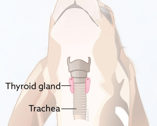
Buddy was diagnosed with thyroid carcinoma. Dogs have two thyroid glands, we only have one! In dogs, they are located at the base of the neck on either side of their windpipe (or trachea).
These glands produce hormones that regulate heart rate, blood pressure, body temperature and weight. Surgical removal of the thyroid gland, when possible, is the recommended treatment for this disease. Buddy was able to participate in 2 clinical trials, both with the goal to improve diagnosis of cancer in our pets!
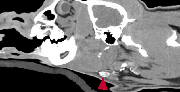
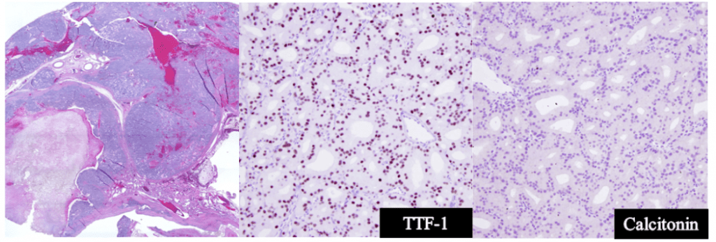
During surgery, Dr Oblak removed his thyroid gland and neighbouring lymph nodes. These tissues were then shipped to Dr Skinner’s team at the Missouri Veterinary Health Centre for a detailed assessment. Any tissues suspect for cancer, typically start with H&E (Hematoxylin and Eosin) staining. By staining the otherwise transparent tissues, we are able to see detailed picture of the tissue including the cellular structures, organization of cells and any abnormalities. Dr Skinner’s team also used special tissue stains, beyond the routine H&E, to help identify any deposits of cancer cells!
These special stains can also help determine the cancer subtype. Thyroid carcinoma has 4 subtypes: follicular, medullary, compact and mixed. This data will be used to assess how and if subtype affects prognosis and the frequency and pattern of thyroid cancer spread to lymph nodes.
These are super magnified images of the thyroid gland tissues. Based on the cellular staining, Buddy has follicular thyroid carcinoma.
Buddy is doing well and was in recently to collect additional blood samples for the study!
