

The OVC Neurology Service consists of three Board Certified Neurologists. They offer a wide variety of treatment services including: hearing tests, spinal decompressive surgery for intervertebral disc disease (IVDD), diagnosis and medical management of epilepsy, management of spinal pain, diagnosis of muscle and nerve disorders, and diagnosis and management of congenital and degenerative nervous system diseases. To learn more about the OVC Neurology Service, please click here.
Understanding Spinal Muscle Function in Dogs with Intervertebral Disc Disorders
Scientific Title: Canine Paraspinal Muscle Biomechanical and Physiological Properties Related to Intervertebral Disc Disorders
Study Investigator: Drs Alex Chan, Fiona James and Stephen Brown
Graduate Student: Josh Briar (PhD)
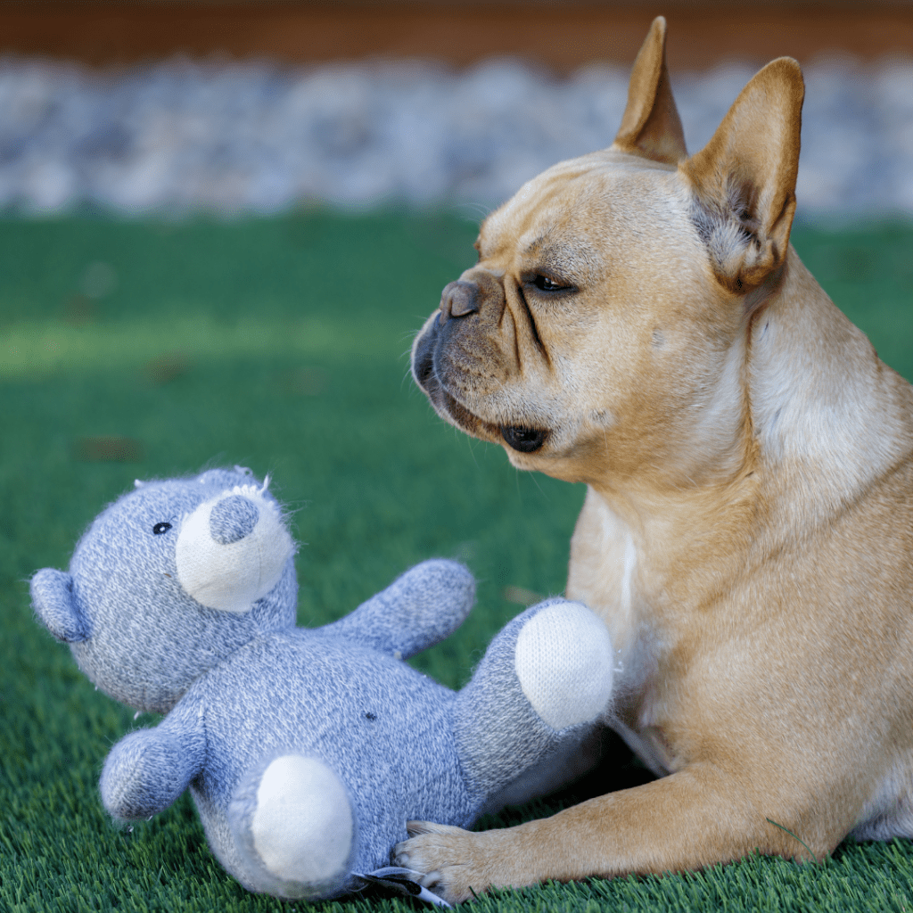
Spinal degeneration causes pain and disability in both dogs and humans with treatment often involving surgery. Because spinal degeneration is associated with the degeneration and dysfunction of spinal muscles, these muscles are a prime target for patient rehabilitation strategies. In humans and dogs, we do not fully understand how these muscles degenerate and the impact of these changes on muscle function. The research team is working closely with human clinicians and recruiting both humans and dogs into this comparative study!
Inclusion criteria:
- Dogs with a confirmed diagnosis of acute disk herniation and/or intervertebral disc degeneration (IVDD) undergoing MRI and standard of care surgery
ON HOLD FOR RECRUITMENT – Investigating Canine Behaviour using Wearable Biomonitors
Scientific Title: Pilot study of wearable biomonitors to explore the behavioural and environmental context of canine seizures
Study Investigator: Dr. Fiona James
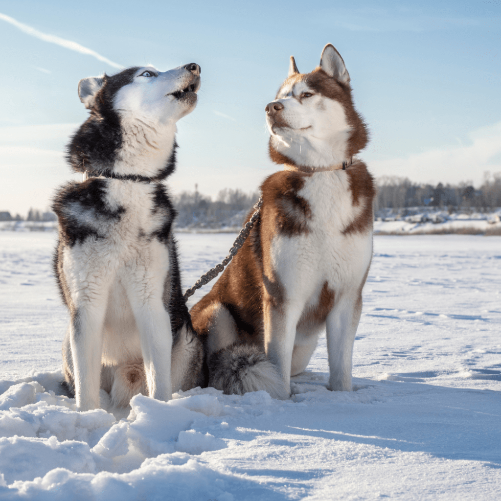
Idiopathic epilepsy (IE) is the most common neurological disorder diagnosed in dogs. IE has a significant negative impact on affected dogs and their owners’ quality of life. Through the use of wearable technologies for both pets and owners, we can increase our understanding of epileptic canine behaviour and gain insight to better predict future seizure occurrence.
Financial incentives are available. This study is fully funded by OVC Pet Trust.
Inclusion criteria:
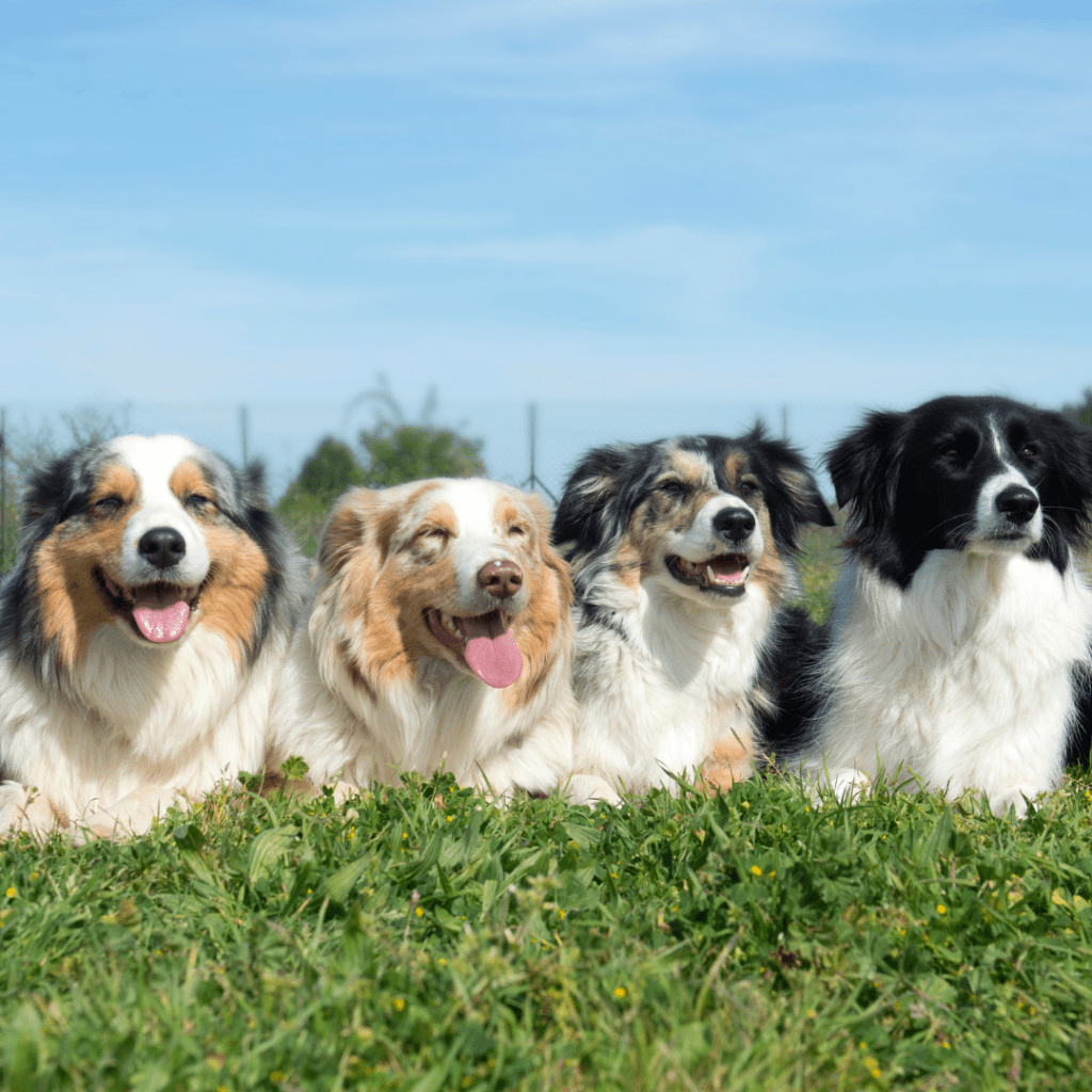
- Dogs (Golden retrievers, Labrador retrievers, Huskies, German Shepherds, Border Collies, Australian Shepherds, Standard Poodles, and mixes of these breeds) between the ages of 2-7 years old
- Two groups:
- Dogs with Tier I idiopathic epilepsy diagnosis with no other health concerns/underlying conditions
- Neurotypical with no other health concerns/underlying conditions
Are These Seizures in Dogs?
Study Investigator: Dr. Fiona James
Graduate Student: Charly McKenna (PhD)
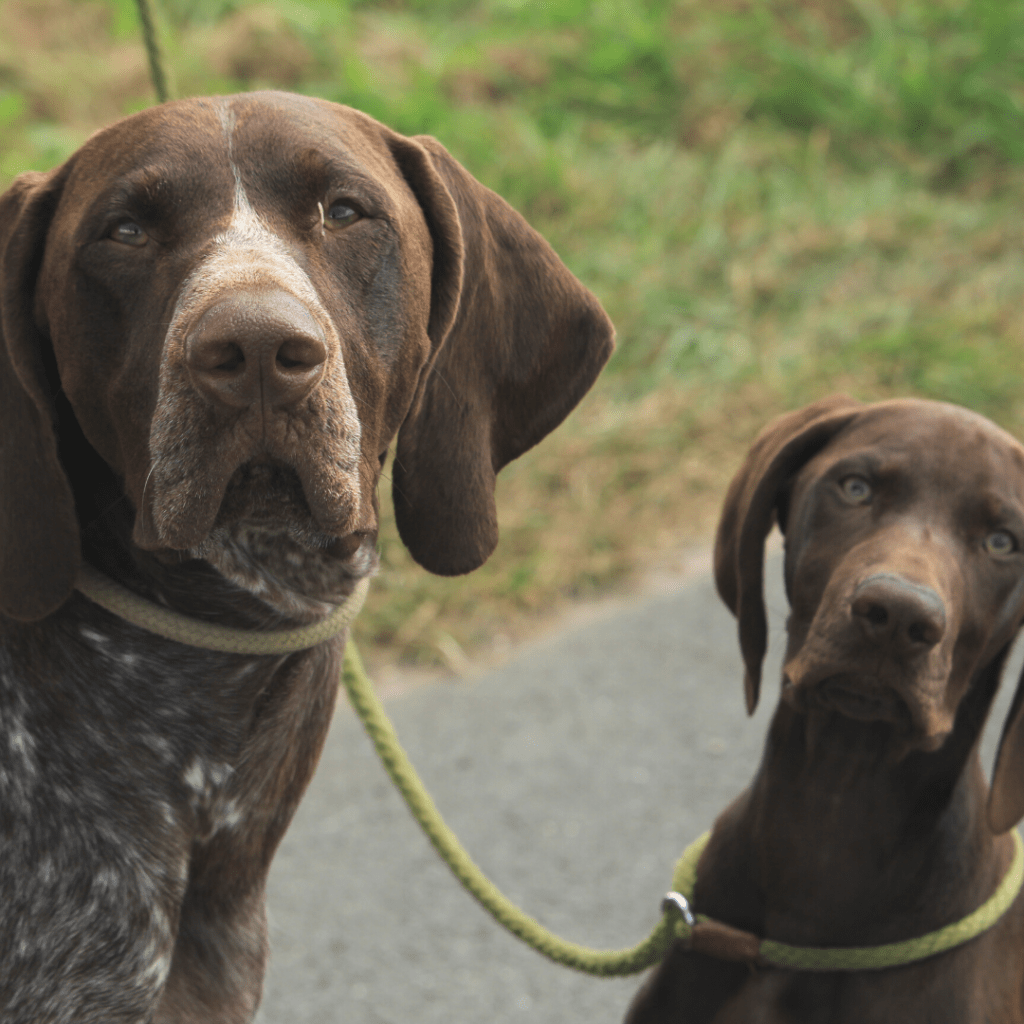
Seizures are common in veterinary medicine, and epilepsy is the most common brain disease in dogs. Diagnosing epilepsy can be tricky because there are many different kinds of seizures and other disorders might look like epilepsy. It’s important to know if a dog’s episodes are actually seizures, as they might need treatment. Using video recording and electroencephalography (EEG), we can confirm if seizure activity is happening and classify the type of seizure.
Financial incentives are available. This study is partially funded by OVC Pet Trust, the American Kennel Club Canine Health Foundation and NSERC.
Inclusion criteria:
- Dogs that are between 6 months and 6 years of age that experience episodes similar to seizures
Do Anti-Seizure Drugs Work in Dogs?
Study Investigator: Dr. Fiona James
Graduate Student: Charly McKenna (PhD)
Even with anti-seizure drug (ASD) treatment, a proportion of dogs may continue to have seizures or experience intolerable side effects from their medication. Accurate seizure control impacts the quality of life and survival in epileptic dogs and also their caretaker’s quality of life.
Financial incentives are available. This study is fully funded by OVC Pet Trust, the American Kennel Club Canine Health Foundation and NSERC.
Inclusion criteria:
- Dogs between 6 months and 6 years of age with normal neurologic exam and at least one year since index seizure (known as Tier I idiopathic epilepsy)
- Any dog with Tier II idiopathic epilepsy regardless of age, neurologic exam status, or time since first onset of seizures (must have normal MRI and CSF & bloodwork)
- Dog’s veterinarian is planning to add a new ASD regardless of previous ASD status
Evaluating the Structural Brain Differences of Dogs Diagnosed with Idiopathic Epilepsy
Scientific Title: Craniocerebral Topographical Mapping in Dogs with Idiopathic Epilepsy
Study Investigator: Dr. Fiona James
Graduate Student: Grace Kadler (PhD)
Diffusion tensor imaging (DTI) is sensitive to the molecular movement of water and can provide information on the integrity and pathology of the brain on a cellular level. In dogs with idiopathic
epilepsy (IE), the microstructures within the brain may be compromised. In order to better understand the abnormal brain structures associated with IE and
potentially improve diagnostic and treatment options, we first need to compare DTI between IE and neurotypical dogs.
Financial incentives are available. This study is partially funded by OVC Pet Trust.
Inclusion criteria:
- All dogs must be mesocephalic (have an average muzzle length) and undergo a MRI at the Ontario Veterinary College
- Neurotypical dogs with no obvious structural brain abnormalities and/or neurological disorders
- Dogs with no physical head abnormalities with a planned EEG recording
- Dogs diagnosed with generalized IE that have no other medical conditions
Investigating the Placement of EEG Electrodes in Dogs with Epilepsy Using 3D Reconstruction
Scientific Title: Craniocerebral Topographical Mapping for Improved Canine Electroencephalographic (EEG) Lesion Localization
Study Investigator: Dr. Fiona James
Graduate Student: Dr. Stephen Everest (DVSc)
To improve the diagnosis and characterization of canine epilepsy, we need to better understand how our scalp electrodes for electroencephalography (EEG) map to the underlying brain surface and establish a best practice for electrode placement and subsequent seizure detection.
Financial incentives are available. This study is partially funded by OVC Pet Trust.
Inclusion criteria:
- All dogs must be mesocephalic (have an average muzzle length) and undergo a MRI and CT at the Ontario Veterinary College
- Neurotypical dogs with no obvious structural brain abnormalities and/or neurological disorders
- Dogs with no physical head abnormalities with a planned EEG recording
- Dogs diagnosed with generalized IE that have no other medical conditions