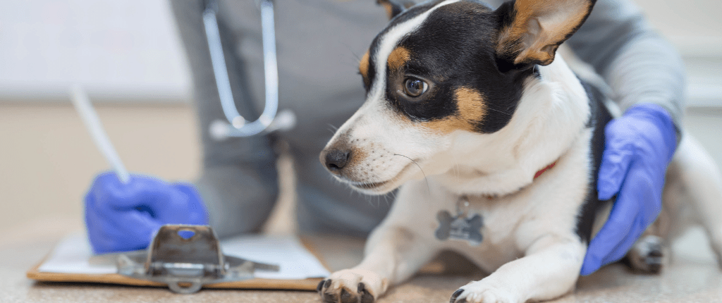
The Companion Animal Surgical Service at the Ontario Veterinary College offers neurosurgery, abdominal and thoracic surgery, cardiovascular and oncologic surgery, and reconstructive surgery. The Surgical Service also offers specialized techniques such as minimally invasive endoscopic surgery, laser procedures and hip replacements. To learn more about the Companion Animal Surgical Service at the OVC, please click here.
Understanding Spinal Muscle Function in Dogs with Intervertebral Disc Disorders
Scientific Title: Canine Paraspinal Muscle Biomechanical and Physiological Properties Related to Intervertebral Disc Disorders
Study Investigator: Drs Alex Chan, Fiona James and Stephen Brown
Graduate Student: Josh Briar (PhD)
Spinal degeneration causes pain and disability in both dogs and humans with treatment often involving surgery. Because spinal degeneration is associated with the degeneration and dysfunction of spinal muscles, these muscles are a prime target for patient rehabilitation strategies. In humans and dogs, we do not fully understand how these muscles degenerate and the impact of these changes on muscle function. The research team is working closely with human clinicians and recruiting both humans and dogs into this comparative study!
Inclusion criteria:
- Dogs with a confirmed diagnosis of acute disk herniation and/or intervertebral disc degeneration (IVDD) undergoing MRI and standard of care surgery
Evaluating the Use of Fluorescent Dyes in Dogs Undergoing Liver Mass Removal Surgery
Scientific Title: Evaluating the Use of Fluorescent Dyes in Dogs Undergoing Liver Mass Removal Surgery
Study Investigator: Dr. Brigitte Brisson
Graduate Student: Dr. Rachel Dobberstein (DVSc)
Dogs are prone to developing liver cancer. In humans, fluorescent dyes are used to identify liver tumours and determine appropriate tissue margins for complete surgical removal. This same technique may be used in dogs with liver tumours and as it does in people, the fluorescence imaging may guide surgeons in determining the required margins to remove the entire tumour.
Inclusion criteria:
- Dogs undergoing an open (laparotomy) abdominal surgery at OVC-HSC
- Dogs diagnosed with primary liver neoplasia or abdominal neoplastic lesions that commonly metastasize to the liver by time of diagnosis e.g. splenic HSA.
- Cancer diagnosis will be suspected based on recent (<14 days) abdominal ultrasound or abdominal CT scan at OVC-HSC

Exploring a Novel Nanoparticle Combined with Light Therapy to Treat Oral Tumours in Dogs
Scientific Title: Exploration of Nanoparticle-Enabled Image Guided Photoablation in Veterinary Patients
Study Investigator: Dr. Michelle Oblak
PORPHYSOME-enabled therapies can have an immediate impact on cancer management providing better patient outcomes. This study will evaluate the potential applications of the novel nanomedicine (PORPHYSOMES) and PDT in veterinary clinical patients with oral tumours.
This project is part of the Veterinary Medical Innovation Platform aligned with Dr Michelle Oblak’s research chair with OVC and Animal Health Partners!
Inclusion criteria:
- Dogs ≤ 60kg, with a confirmed oral tumour (any type) and interested in pursuing CT and surgery will be eligible for this study
- There will be two arms: PDT + surgery and monotherapy; these will be explained by the clinical trials team at enrollment
Treatment of Elbow Dysplasia in Dogs with Canine Stem Cells
Scientific Title: Treatment of Elbow Dysplasia Patients with Canine Adipose Tissue-Derived Mesenchymal Stromal Cells
Study Investigator: Dr. Melissa MacIver
Graduate Student: Dr. Rebecca Beardall (DVSc)
Joint inflammation and associated diseases are common in dogs, with around 20% of dogs developing osteoarthritis (OA). Large to giant breed dogs are often diagnosed with elbow dysplasia (ED), which can lead to OA later in life. Current treatments, such as medications (non-steroidal anti-inflammatories (NSAIDs)) and surgery, might not work for all dogs. Mesenchymal stromal cells (MSCs) have shown anti-inflammatory effects and could be a treatment option for immune and inflammatory disorders in dogs, like ED. This alternative may have better safety and efficacy and require less owner compliance compared to NSAIDs.
Inclusion criteria:
- Dogs that have a confirmed diagnosis of bilateral elbow dysplasia and are otherwise healthy
Exploring a Novel Diagnostic and Treatment Technique in Combination With Surgery for Thyroid Tumours in Dogs
Scientific Title: Exploration of Nanoparticle-Enabled Image Guided Photoablation in Veterinary Patients
Study Investigator: Dr. Michelle Oblak
PORPHYSOME-enabled therapies can have an immediate impact on cancer management providing better patient outcomes. This study will evaluate the potential applications of the novel nanomedicine (PORPHYSOMES) and PDT in veterinary clinical patients with thyroid tumours.
This project is part of the Veterinary Medical Innovation Platform aligned with Dr Michelle Oblak’s research chair with OVC and Animal Health Partners!
Inclusion criteria:
- Dogs with a confirmed freely moveable thyroid tumour interested in pursuing surgery are eligible
ON HOLD FOR RECRUITMENT – Evaluation of a Fluorescent dye in Dogs with Hepatobiliary Disease Undergoing Gallbladder Removal Surgery
Scientific Title: Evaluation of Indocyanine Green (ICG) Cholangiography in Dogs with Hepatobiliary Disease Undergoing Cholecystectomy
Study Investigator: Dr. Brigitte Brisson
This study aims to determine the clinical usefulness of a safe fluorescent dye (indocyanine green, ICG) in canine patients with hepatobiliary disease scheduled to undergo gall bladder removal surgery by assessing whether it improves visualization of the biliary tree during surgery.
Inclusion Criteria:
- Any dog undergoing routine gall bladder removal surgery (open or laparoscopic cholecystectomy) during normal operating hours at the OVC
Evaluating the Use of Fluorescence Dyes in Surgery for Dogs with Pancreatic Cancer
Scientific Title: Pilot Evaluation of Near Infrared Fluorescence Imaging for Intraoperative Identification of Canine Insulinoma and Their Metastasis
Study Investigator: Dr. Michelle Oblak
Making tumours glow-in-the-dark, using a combination of near-infrared fluorescence imaging (NIRF) and a fluorescent dye, Indocyanine Green (ICG), has been used in several veterinary applications – many in surgical oncology clinical trials at OVC! During surgery, pancreatic masses in dogs can be challenging to remove as they are often quite small and difficult to see on gross visualization or via traditional imaging methods. Using NIRF-ICG for surgical guidance has the potential to improve patient outcomes and enable better visualization of the pancreatic mass(es) and metastatic sites.
Inclusion criteria:
- Dogs that are diagnosed with a pancreatic mass and scheduled for exploratory laparotomy
The Use of Fluorescent Dyes to Evaluate Sentinel Lymph Nodes During Surgery for Dogs with Lung Tumours
Scientific Title: Evaluation of Sentinel Lymph Node Mapping in Dogs with Lung Tumours using CT Lymphography and Intraoperative Indocyanine Green
Study Investigator: Dr. Michelle Oblak
By developing new protocols, we can ensure accurate evaluation of the most important lymph node(s) to make better follow-up and treatment recommendations. This will help to improve patient treatments and outcomes for dogs diagnosed with lung tumours, as well as dogs and cats with other solid tumour types in the future. The team is working closely with human surgeons on this translational project.
Inclusion criteria:
- Dogs with a single lung tumour (less than 5cm) interested in pursuing CT scan and surgery

Comparison of Two Surgical Techniques and Long Term Outcomes to Alleviate Congenital Constriction in Dogs
Scientific Title: Prospective, Long-Term Evaluation of Esophageal Function and Clinical Outcome Following Surgical Treatment of Vascular Ring Anomalies (VRA) in Dogs
Study Investigator: Dr. Ameet Singh
Vascular ring anomalies (VRA) are a result of
developmental abnormalities during fetal growth. Early surgical treatment of VRA is recommended to alleviate
the clinical signs and prevent long-term abnormalities
to the neuromuscular function of the esophagus. The objective of this study is to evaluate the clinical outcome and esophageal function following surgical treatment of VRA with traditional and minimally invasive techniques.
Inclusion criteria:
- Dogs with a confirmed diagnosis of a VRA undergoing CT scan and surgery
Evaluating the Use of Minimally Invasive Surgical Techniques for Dogs with Spontaneous Pneumothorax
Scientific Title: Prospective Evaluation of Minimally Invasive Thoracic Surgery for Identification of Pulmonary Bullae in Dogs with Spontaneous Pneumothorax
Study Investigator: Dr. Ameet Singh
Pneumothorax is the abnormal presence of free air within the chest cavity outside of the lungs. This can be a life-threatening condition as this air restricts the lungs from inflating normally during inhalation. Currently in veterinary patients, standard of care surgical treatment involves a thorough exploration of the thoracic cavity through the breastbone. Minimally invasive (MI) thoracic surgery may be an alternative approach given the reduction in postoperative pain associated with this technique. While the MI surgery has its advantages, the accuracy in identifying bubbles on the lungs during this technique has not yet been proven.
Inclusion criteria:
- Dogs with spontaneous pneumothorax (not caused by traumatic event or underlying condition) and interested in pursuing surgery
Analyzing Lymph Node Spread in Dogs Undergoing Surgery for Thyroid Tumours
Scientific Title: Evaluation of Regional Lymph Node Metastasis in Canine Thyroid Carcinoma
Study Investigator: Dr. Michelle Oblak
Thyroid tumours are relatively uncommon in dogs, however about 90% of these tumours are either malignant carcinomas or adenocarcinomas. Using special stains, tissues once surgically removed can be categorized into subtypes: follicular, medullary, compact and mixed. The subtype may affect prognosis including the frequency and pattern of thyroid tumours to spread to lymph nodes.
Inclusion criteria:
- Dogs with a confirmed diagnosis of a thyroid tumour undergoing staging and surgery
Comparing the Use of Fluorescent Dyes in Surgery to Previously Reported Methods for Improving Lymph Node Staging in Dogs with Oral Cancer
Scientific Title: Evaluation of Agreement Between Computed Tomography Lymphangiography and The Combination of Methylene Blue and Indocyanine Green for Sentinel Lymph Node Mapping in Dogs with Oral Tumours
Study Investigator: Dr. Michelle Oblak
Development of imaging and intraoperative protocols could help decrease the number of lymph nodes surgically removed, in addition to ensuring accurate evaluation of the most important lymph node(s) for making follow-up treatment recommendations improving patient prognosis and outcomes for dogs diagnosed with oral tumours.
Inclusion criteria:
- Dogs with a diagnosed oral tumour and interested in pursuing CT & Surgery
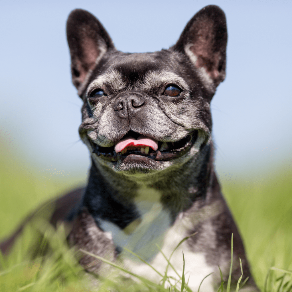
Comparison of Two Surgical Techniques For The Treatment of Brachycephalic Airway Disease in French Bulldogs
Scientific Title: Prospective, Randomized Trial Comparing Two Surgical Techniques For The Treatment of Brachycephalic Obstructive Airway Syndrome (BOAS) in French Bulldogs
Study Investigator: Dr. Ameet Singh
Currently in veterinary patients, standard of care surgery for BOAS is a soft palate resection (staphylectomy). Given that some dogs still suffer from breathing difficulties following surgery, modifications that aim to provide a greater opening of the airway have been proposed. One of these procedures is the folded flap palatoplasty however a comparison of these techniques as it relates to clinical outcome has not been performed.
Inclusion criteria:
- French bulldogs <5 years of age, with breathing difficulties as a result of BOAS and are interested in pursuing CT and surgery

Evaluating the Use of Fluorescent Dyes to Assess Blood Flow in Dogs Undergoing Intestinal Resection During Foreign Body Surgery
Scientific Title: Use of Fluorescent Dyes for Perfusion Assessment and Surgical Planning for Foreign Body Surgery in Dogs
Study Investigator: Dr. Michelle Oblak
Graduate Student: Dr. Rey Zhang (DVSc)
In human medicine, the use of fluorescent dyes like Indocyanine Green (ICG), have been found to decrease complications in bowel surgery. ICG has not yet been used for intestinal blood flow assessment in pets but might be helpful to reduce complications associated with poor blood flow including poor healing and leakage.
Inclusion Criteria:
- Dogs undergoing intestinal surgery for foreign body removal that require part of their intestines


